Predicting preoperative lymph node metastasis of hilar cholangiocarcinoma based on deep learning radiomics
Keywords:
Hilar cholangiocarcinoma, Deep learning, Transfer learning, UltrasoundAbstract
Background: The objective of this study was to evaluate whether deep learning radiomic (DLR) models utilizing B-mode ultrasound (BUS) and contrast-enhanced ultrasound (CEUS) could improve the preoperative prediction of lymph node metastasis (LNM) in patients with hilar cholangiocarcinoma (HCCA).
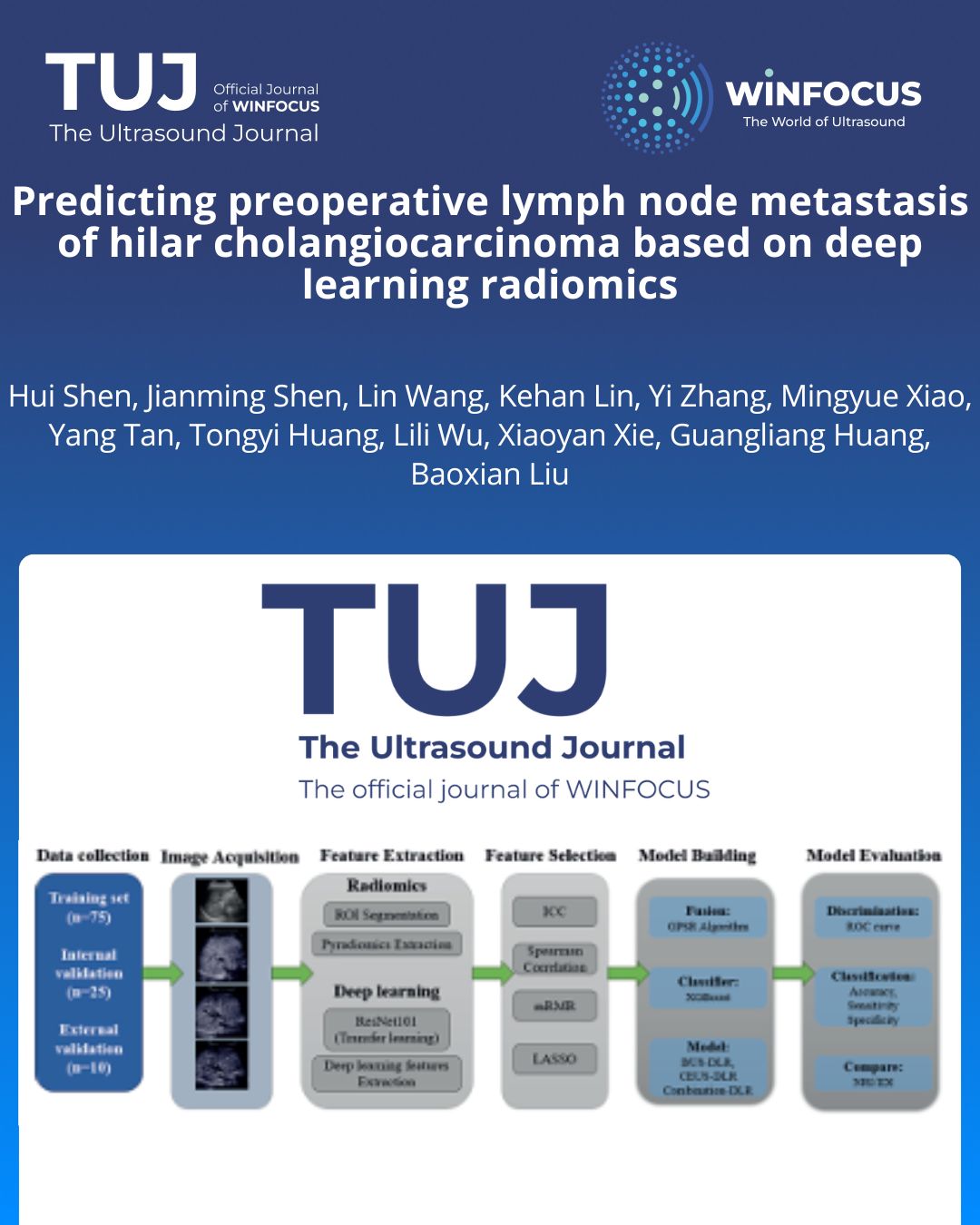
Methods: The study included 110 HCCA patients from two clinical centers, divided into primary and external validation cohorts. Pathological verification of lymph node status was performed, and the ResNet101 architecture was used to extract deep learning features (DLFs) from BUS and CEUS images. The Genetic Programming-based Symbolic Regression (GPSR) algorithm was applied to integrate radiomic features (RadFs) and DLFs, generating deep learning radiomic features (DLRFs). DLR models were subsequently constructed using the eXtreme Gradient Boosting (XGBoost) algorithm.
Results: Lymph node metastasis was identified in 48 out of 110 patients (43.64%). No significant differences in clinical characteristics were observed between LNM-positive and LNM-negative groups (P-values ranging from 0.14 to 0.98). A total of 837 RadFs and 4095 DLFs were initially extracted from each tumor region of interest (ROI). After feature selection, 10 RadFs (4 from BUS, 6 from CEUS) and 27 DLFs (5 from BUS, 22 from CEUS) were retained. Using the GPSR algorithm, 5 BUS-DLRFs, 10 CEUS-DLRFs, and 15 Combination-DLRFs were generated, leading to the development of three corresponding DLR models. In internal validation, the AUC values were 0.70 for the BUS-DLR model, 0.77 for the CEUS-DLR model, and 0.83 for the Combination-DLR model. In external validation, the AUC values were 0.66, 0.68, and 0.72, respectively. These results indicate that the integration of multiphasic CEUS and BUS data is essential for more comprehensively identifying LNM and achieving precise preoperative staging.
Conclusions: The DLR models based on DLRFs demonstrated an enhanced ability to preoperatively predict LNM in HCCA patients, indicating that the integration of deep learning and RadFs from BUS and CEUS may offer improved predictive performance for LNM.
References
1. Bergquist A, von Seth E. Epidemiology of cholangiocarcinoma. Best Pract Res Clin Gastroenterol. 2015;29(2):221-32. doi:10.1016/j.bpg.2015.02.003
2. DeSantis CE, Kramer JL, Jemal A. The burden of rare cancers in the United States. CA Cancer J Clin. 2017;67(4):261-72. doi:10.3322/caac.21400
3. Li B, Li Z, Qiu Z, Qin Y, Gao Q, Ao J, et al. Surgical treatment of hilar cholangiocarcinoma: retrospective analysis. BJS Open. 2023;7(3). doi:10.1093/bjsopen/zrad024
4. Sapisochin G, Ivanics T, Subramanian V, Doyle M, Heimbach JK, Hong JC. Multidisciplinary treatment for hilar and intrahepatic cholangiocarcinoma: A review of the general principles. Int J Surg. 2020;82s:77-81. doi:10.1016/j.ijsu.2020.04.067
5. Ito F, Cho CS, Rikkers LF, Weber SM. Hilar cholangiocarcinoma: current management. Ann Surg. 2009;250(2):210-8. doi:10.1097/SLA.0b013e3181afe0ab
6. Banales JM, Marin JJG, Lamarca A, Rodrigues PM, Khan SA, Roberts LR, et al. Cholangiocarcinoma 2020: the next horizon in mechanisms and management. Nat Rev Gastroenterol Hepatol. 2020;17(9):557-88. doi:10.1038/s41575-020-0310-z
7. Liu ZP, Zhang QY, Chen WY, Huang YY, Zhang YQ, Gong Y, et al. Evaluation of Four Lymph Node Classifications for the Prediction of Survival in Hilar Cholangiocarcinoma. J Gastrointest Surg. 2022;26(5):1030-40. doi:10.1007/s11605-021-05211-x
8. Hau HM, Meyer F, Jahn N, Rademacher S, Sucher R, Seehofer D. Prognostic Relevance of the Eighth Edition of TNM Classification for Resected Perihilar Cholangiocarcinoma. J Clin Med. 2020;9(10). doi:10.3390/jcm9103152
9. Gaspersz MP, Buettner S, van Vugt JLA, de Jonge J, Polak WG, Doukas M, et al. Evaluation of the New American Joint Committee on Cancer Staging Manual 8th Edition for Perihilar Cholangiocarcinoma. J Gastrointest Surg. 2020;24(7):1612-8. doi:10.1007/s11605-019-04127-x
10. Buettner S, van Vugt JLA, Gaspersz MP, Coelen RJS, Roos E, Labeur TA, et al. Survival after resection of perihilar cholangiocarcinoma in patients with lymph node metastases. HPB (Oxford). 2017;19(8):735-40. doi:10.1016/j.hpb.2017.04.014
11. Groot Koerkamp B, Wiggers JK, Allen PJ, Besselink MG, Blumgart LH, Busch OR, et al. Recurrence Rate and Pattern of Perihilar Cholangiocarcinoma after Curative Intent Resection. J Am Coll Surg. 2015;221(6):1041-9. doi:10.1016/j.jamcollsurg.2015.09.005
12. Guglielmi A, Ruzzenente A, Campagnaro T, Pachera S, Conci S, Valdegamberi A, et al. Prognostic significance of lymph node ratio after resection of peri-hilar cholangiocarcinoma. HPB (Oxford). 2011;13(4):240-5. doi:10.1111/j.1477-2574.2010.00277.x
13. Mao K, Liu J, Sun J, Zhang J, Chen J, Pawlik TM, et al. Patterns and prognostic value of lymph node dissection for resected perihilar cholangiocarcinoma. J Gastroenterol Hepatol. 2016;31(2):417-26. doi:10.1111/jgh.13072
14. Choi JY, Kim MJ, Lee JM, Kim KW, Lee JY, Han JK, et al. Hilar cholangiocarcinoma: role of preoperative imaging with sonography, MDCT, MRI, and direct cholangiography. AJR Am J Roentgenol. 2008;191(5):1448-57. doi:10.2214/ajr.07.3992
15. Lee HY, Kim SH, Lee JM, Kim SW, Jang JY, Han JK, et al. Preoperative assessment of resectability of hepatic hilar cholangiocarcinoma: combined CT and cholangiography with revised criteria. Radiology. 2006;239(1):113-21. doi:10.1148/radiol.2383050419
16. Akamatsu N, Sugawara Y, Osada H, Okada T, Itoyama S, Komagome M, et al. Diagnostic accuracy of multidetector-row computed tomography for hilar cholangiocarcinoma. J Gastroenterol Hepatol. 2010;25(4):731-7. doi:10.1111/j.1440-1746.2009.06113.x
17. Gu XJ, Wang BF, Liu R. [Application of ¹⁸F-fluorodeoxyglucose positron emission tomography/computed tomography in preoperative assessment of hilar cholangiocarcinoma]. Zhonghua Yi Xue Za Zhi. 2012;92(20):1409-12. PMID: 22883200
18. Nielsen MB, Søgaard SB, Bech Andersen S, Skjoldbye B, Hansen KL, Rafaelsen S, et al. Highlights of the development in ultrasound during the last 70 years: A historical review. Acta Radiol. 2021;62(11):1499-514. doi:10.1177/02841851211050859
19. Postema M, Gilja OH. Contrast-enhanced and targeted ultrasound. World J Gastroenterol. 2011;17(1):28-41. doi:10.3748/wjg.v17.i1.28
20. du Bois H, Heim TA, Lund AW. Tumor-draining lymph nodes: At the crossroads of metastasis and immunity. Sci Immunol. 2021;6(63):eabg3551. doi:10.1126/sciimmunol.abg3551
21. Avraamides CJ, Garmy-Susini B, Varner JA. Integrins in angiogenesis and lymphangiogenesis. Nat Rev Cancer. 2008;8(8):604-17. doi:10.1038/nrc2353
22. Kloth C, Kratzer W, Schmidberger J, Beer M, Clevert DA, Graeter T. Ultrasound 2020 - Diagnostics & Therapy: On the Way to Multimodal Ultrasound: Contrast-Enhanced Ultrasound (CEUS), Microvascular Doppler Techniques, Fusion Imaging, Sonoelastography, Interventional Sonography. Rofo. 2021;193(1):23-32. doi:10.1055/a-1217-7400
23. Zheng X, Yao Z, Huang Y, Yu Y, Wang Y, Liu Y, et al. Deep learning radiomics can predict axillary lymph node status in early-stage breast cancer. Nat Commun. 2020;11(1):1236. doi:10.1038/s41467-020-15027-z
24. Yu J, Deng Y, Liu T, Zhou J, Jia X, Xiao T, et al. Lymph node metastasis prediction of papillary thyroid carcinoma based on transfer learning radiomics. Nat Commun. 2020;11(1):4807. doi:10.1038/s41467-020-18497-3
25. Tao L, Zhou W, Zhan W, Li W, Wang Y, Fan J. Preoperative Prediction of Cervical Lymph Node Metastasis in Papillary Thyroid Carcinoma via Conventional and Contrast-Enhanced Ultrasound. J Ultrasound Med. 2020;39(10):2071-80. doi:10.1002/jum.15315
26. Huang YQ, Liang CH, He L, Tian J, Liang CS, Chen X, et al. Development and Validation of a Radiomics Nomogram for Preoperative Prediction of Lymph Node Metastasis in Colorectal Cancer. J Clin Oncol. 2016;34(18):2157-64. doi:10.1200/jco.2015.65.9128
27. Bera K, Braman N, Gupta A, Velcheti V, Madabhushi A. Predicting cancer outcomes with radiomics and artificial intelligence in radiology. Nat Rev Clin Oncol. 2022;19(2):132-46. doi:10.1038/s41571-021-00560-7
28. Xu X, Zhang HL, Liu QP, Sun SW, Zhang J, Zhu FP, et al. Radiomic analysis of contrast-enhanced CT predicts microvascular invasion and outcome in hepatocellular carcinoma. J Hepatol. 2019;70(6):1133-44. doi:10.1016/j.jhep.2019.02.023
29. Gillies RJ, Kinahan PE, Hricak H. Radiomics: Images Are More than Pictures, They Are Data. Radiology. 2016;278(2):563-77. doi:10.1148/radiol.2015151169
30. Tang X, Zhuang Z, Jiang L, Zhu H, Wang D, Zhang L. A Preoperative CT-based Multiparameter Deep Learning and Radiomic Model with Extracellular Volume Parameter Images Can Predict the Tumor Budding Grade in Rectal Cancer Patients. Academic radiology. 2025;32(7):4002-12. doi:10.1016/j.acra.2025.02.028
31. Yin S, Ding N, Ji Y, Qiao Z, Yuan J, Chi J, et al. The value of CT radiomics combined with deep transfer learning in predicting the nature of gallbladder polypoid lesions. Acta Radiol. 2024;65(6):554-64. doi:10.1177/02841851241245970
32. Liu R, Yuan F, Wang B, Chen W, Ye J, He Y. A novel deep learning model based on multimodal contrast-enhanced ultrasound dynamic video for predicting occult lymph node metastasis in papillary thyroid carcinoma. Front Endocrinol (Lausanne). 2025;16:1634875. doi:10.3389/fendo.2025.1634875
33. Liu F, Liu D, Wang K, Xie X, Su L, Kuang M, et al. Deep Learning Radiomics Based on Contrast-Enhanced Ultrasound Might Optimize Curative Treatments for Very-Early or Early-Stage Hepatocellular Carcinoma Patients. Liver cancer. 2020;9(4):397-413. doi:10.1159/000505694
34. Liu D, Yang K, Zhang C, Hu Z, Cheng Y, Liu Y. Deep Learning with Contrast-Enhanced Ultrasound for Preoperative Prediction of Early Recurrence in Hepatocellular Carcinoma After Hepatectomy. J Multidiscip Healthc. 2025;18:6829-41. doi:10.2147/jmdh.S555110
35. Hu H-t, Shan Q-y, Chen S-l, Li B, Feng S-t, Xu E-j, et al. CT-based radiomics for preoperative prediction of early recurrent hepatocellular carcinoma: technical reproducibility of acquisition and scanners. La Radiologia medica. 2020;125(8):697-705. doi:10.1007/s11547-020-01174-2
36. Wang Y, Shao J, Wang P, Chen L, Ying M, Chai S, et al. Deep Learning Radiomics to Predict Regional Lymph Node Staging for Hilar Cholangiocarcinoma. Front Oncol. 2021;11:721460. doi:10.3389/fonc.2021.721460
37. Hosokawa I, Hayano K, Furukawa K, Takayashiki T, Kuboki S, Takano S, et al. Preoperative Diagnosis of Lymph Node Metastasis of Perihilar Cholangiocarcinoma Using Diffusion-Weighted Magnetic Resonance Imaging. Ann Surg Oncol. 2022;29(9):5502-10. doi:10.1245/s10434-022-11931-4
38. Kim HE, Cosa-Linan A, Santhanam N, Jannesari M, Maros ME, Ganslandt T. Transfer learning for medical image classification: a literature review. BMC Med Imaging. 2022;22(1):69. doi:10.1186/s12880-022-00793-7
39. Zahid U, Ashraf I, Khan MA, Alhaisoni M, Yahya KM, Hussein HS, et al. BrainNet: Optimal Deep Learning Feature Fusion for Brain Tumor Classification. Comput Intell Neurosci. 2022;2022:1465173. doi:10.1155/2022/1465173
40. Wang W, Chen LD, Lu MD, Liu GJ, Shen SL, Xu ZF, et al. Contrast-enhanced ultrasound features of histologically proven focal nodular hyperplasia: diagnostic performance compared with contrast-enhanced CT. Eur Radiol. 2013;23(9):2546-54. doi:10.1007/s00330-013-2849-3
41. Dietrich CF, Nolsøe CP, Barr RG, Berzigotti A, Burns PN, Cantisani V, et al. Guidelines and Good Clinical Practice Recommendations for Contrast-Enhanced Ultrasound (CEUS) in the Liver-Update 2020 WFUMB in Cooperation with EFSUMB, AFSUMB, AIUM, and FLAUS. Ultrasound Med Biol. 2020;46(10):2579-604. doi:10.1016/j.ultrasmedbio.2020.04.030
42. Soares KC, Jarnagin WR. The Landmark Series: Hilar Cholangiocarcinoma. Ann Surg Oncol. 2021;28(8):4158-70. doi:10.1245/s10434-021-09871-6
Downloads
Additional Files
Published
Issue
Section
License
Copyright (c) 2026 Hui Shen, Jianming Shen, Lin Wang, Kehan Lin, Yi Zhang, Mingyue Xiao, Yang Tan, Tongyi Huang, Lili Wu, Xiaoyan Xie, Guangliang Huang, Baoxian Liu (Author)

This work is licensed under a Creative Commons Attribution-NonCommercial 4.0 International License.
This is an Open Access article distributed under the terms of the Creative Commons Attribution License (https://creativecommons.org/licenses/by-nc/4.0) which permits unrestricted use, distribution, and reproduction in any medium, provided the original work is properly cited.
Authors retain the copyright for their published work. No formal permission will be required to reproduce parts (tables or illustrations) of published papers, provided the source is quoted appropriately and reproduction has no commercial intent.
How to Cite